Echinoderma
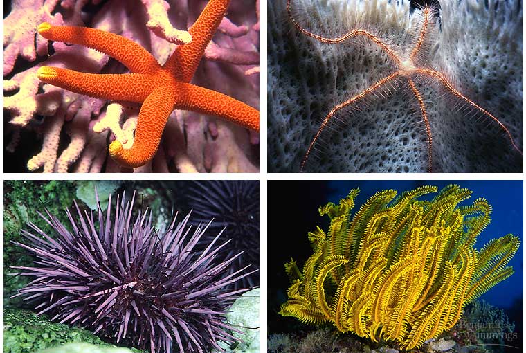
Echinoderma or the sea stars and their relatives consists of about 6,000 living species, all of which are marine. The characteristics that distinguish Phylum Echinodermata are radial symmetry, an internal skeleton, and a water-vascular system. Although adult echinoderms are radially symmetrical, they start out life as a bilateral larvae and they evolved from a bilaterally symmetrical ancestor.
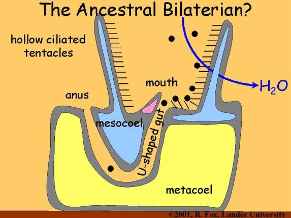
Fossils inform us that the first echinoderms were sessile, feeding by "arms" that surrounded an upward-turned mouth. Since radially symmetry is an effective body plan for this type of life style, the bilaterally symmetrical larvae of early echinoderms became transformed into a radially symmetrical shape during metamorphosis.
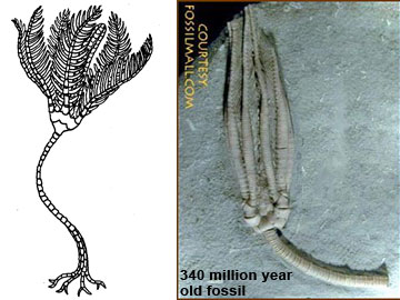
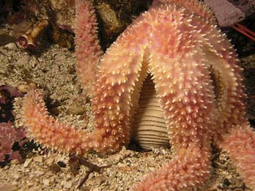
A few modern echinoderms have retained this ancient, sessile life style, but most are freely moving hunters or grazers.
The body plan of echinoderms is designed to utilize water pressure for locomotion. This water vascular system is unique to echinoderms and accounts for much of their success as a group.
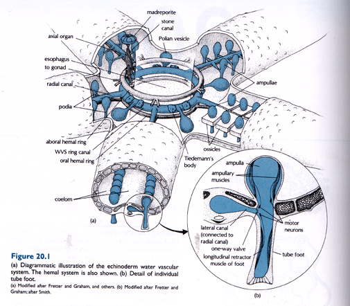
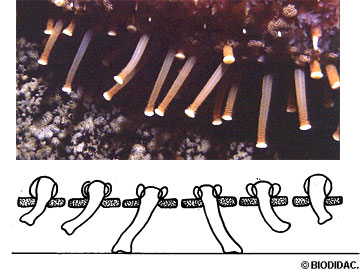
Water enters the water vascular system through a sieve plate and passes into radial canals within each arm.
Official pathway according to your book: aboral madreporite --> stone canal --> ring canal --> 5 radial canals --> numerous am pull ae bearing tube feet.

In summary, numerous cross connecting canals within each arm are attached to hollow structures called tube feet. Small muscles in conjunction with water pressure can extend, contract, and move the tube feet to provide a type of walking locomotion. Longitudinal musculature of the ampulla contracts and the valve in the lateral canal closes, forcing water into the tube foot. When the tube foot contacts the substratum, the sucker adheres. Biologists long thought that a suction disc at the end of each tube foot provided all the animal’s holding power. However, recent investigations revealed that the tube foot can supplement its disc suction with a chemical cement that significantly increases holding power. As the sea star moves, only a subset of its many tube feet contact the ground at any one time.
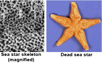
Echinoderms have an internal skeleton made of bony plates (ossicles) of calcium carbonate. They deposit this material after extracting dissolved calcium and carbonate (bicarbonate) ions from sea water. In some species, such as the sea urchin, plates of the skeleton are locked together to form a rigid structure. On the other hand, most sea stars and brittle stars can flex their ‘arms’, indicating that the skeleton has gaps and flexible plate junctures. Sea cucumbers have no real skeleton; only tiny remnant ossicles.
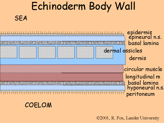
In most echinoderms, spines extend from the skin, accounting for the phylum name which means "spiny-skinned" animals. The spines also are part of the internal skeleton and are covered by epidermis.

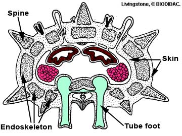
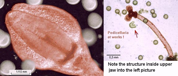
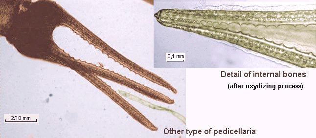
A unique and amazing feature of the echinoderm skin is the presence of tiny, jaw-like structures, pedicellariae, that cover the upper surface of the body. They can open and close to trap tiny animals. Mainly used to defend the aboral surface against settling larvae or other small animals. They consist of a short, fleshy stalk ending in two but sometimes three, small movable ossicles arranged to form grasping jaws.
A more jaw like pedicellariae
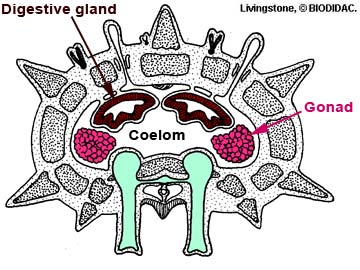
Most internal organs of echinoderms are housed within a large body cavity, the coelom, and are distributed symmetrically around the central body axis. Echinoderms are said to have pentamerous radial symmetry, since the body of most species is divided into five similar parts.
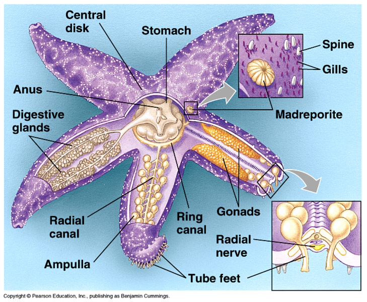
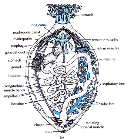
Aside from the water vascular system, designed primarily for locomotion, the organ systems of the echinoderms are quite simple. The water vascular system and the fluid with the coelom circulate oxygen to throughout body, and nutrients also distributed by diffusion through the coelom. There is no excretory system: metabolic wastes simply diffuse out through the skin or tube feet. Sea stars have small out-pocketings of skin, papulae, that extend through the endoskeleton to increase the area for respiration, whereas sea urchins utilize their long tube feet for this purpose. The sand dollar has specialized tube feet that extend from the dorsal surface to absorb oxygen from the water. The sea cucumbers are unusual. They have developed a "respiratory tree" that takes in water through the anus and functions like an internal gill.
.
There are specific organs associated with digestion and nutrient absorption. In sea stars, where digestion is often external to the body, the digestive system consists of a large stomach, but a very short intestine. Digestive glands (pyloric caeca) secrete digestive enzymes into the cardiac stomach, and digested food then passes into the digestive glands where it is absorbed and stored. The small amount of undigested food is eliminated through a dorsal anus. In sea stars the stomach is eversible.
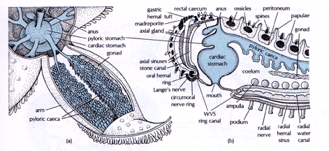
The pathway: cardiac stomach (eversible)-->pyloric stomach-->paired digestive glands (pyloric cecae) in each arm.
Animals that feed on plant material or extract their food from sediments usually have a longer digestive tract. This is true of the sea urchins and sea cucumbers, which have a very long intestine. Some sea urchins and cucumbers also have Polian vesicles that contain excess fluid and ameobocytes. Ameobocytes do exchange materials with surrounding tissue.
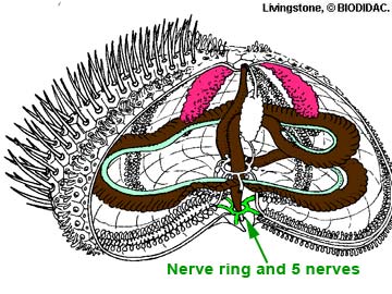
Echinoderms have a simple nervous system consisting of a central nerve ring and nerves that lead to the periphery. This provides a limited amount of coordination so that the animal can move with one part of the body leading. Sensory structures consist mainly of scattered cells sensitive to chemicals and touch, although sea stars also have light-sensitive eye spots at the tips of their arms and lately there is some indication that the general dorsal surface may be sensitive to light.
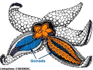
There is a hemal system or "circulatory" system, although its function is somewhat unknown. The spongy hemal system parallels the water vascular system and hemal channels extend to the gonads. Hemal system may function in transporting nutrients from the coelomic fluid to the gonads. With labeled food material, radioactivity appeared in the digestive system, hemal system and finally in the gonads.
Almost all echinoderms have separate sexes and reproduce by shedding eggs and sperm into the water.
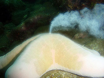
This type of external fertilization is typical of marine organisms. There is usually a way of assuring that many animals spawn at the same time in the same place to produce a large number of offspring. In sea stars, the timing of reproduction is determined by day length. Thus all animals have mature gonads at approximately the same time. When the first sea star spawns, released chemicals cause spawning in all nearby sea stars of the same species, assuring that sperm come in contact with eggs.
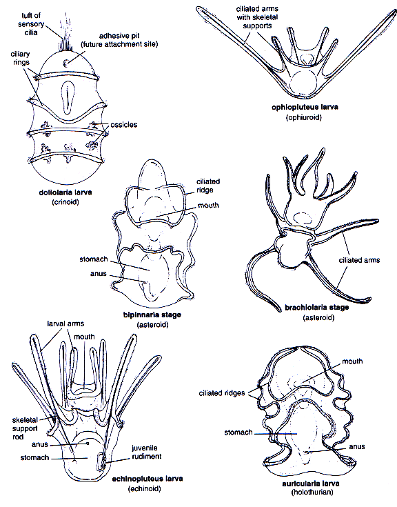
Fertile eggs of echinoderms develop into tiny, swimming larvae that are bilaterally symmetrical. After a few weeks, the larvae attach to a solid substrate and undergo metamorphosis into the adult body form.
.
Sep 11 2003 - Physiology Web Site
Young Sea Animals Clone Themselves—century-old Debate Halted
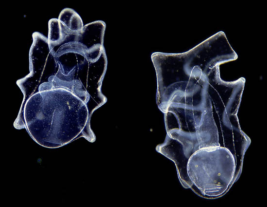
After more than a century of intensive study, scientists have assumed that larvae of non-parasitic invertebrates reproduce only very rarely, but new research by University of Alberta scientists overthrows this conventional wisdom. Graduate student Alexandra Eaves and Dr. Richard Palmer, from the U of A’s Faculty of Science, have found that asexual cloning by some marine invertebrate larvae is not as rare and enigmatic a phenomenon as previously assumed.
“A wealth of knowledge of how embryos grow has come from studying sea urchin development,” said Eaves. “The discovery that these young animals can clone themselves provides an exceedingly rare opportunity to examine how a growing animal can repeat its own early development using a part of its body.”
Scattered earlier reports have observed that invertebrate larvae can spontaneously clone but Eaves and Palmer discovered this trait in three new echinoderm groups–sea cucumbers, sand dollars, and sea urchins–offering surprising new insight about chordate evolution. Larval cloning represents an intriguing new dimension to invertebrate life histories including the suggestion that clones may subsequently clone. The research is published in the current edition of the prestigious journal Nature.
The larvae of echinoderms (the group that includes starfish, sea urchins, sea cucumbers, etc.) usually swim and feed for several months before transforming into a miniature adult. During this time some larvae form an outgrowth – essentially a ball of tissue – that pinches-off of the larval body and grows into a second, normal-looking larva–a clone.
One of the most remarkable parts about the research is that for more than 100 years, scientists may have observed larval asexual reproduction, but did not recognize what they saw, said Palmer. Even more remarkably, at least one early report of larval cloning was dismissed as an artifact of laboratory culture conditions.
“These data make it clear that people have likely seen this spontaneous cloning for many years but not recognized it,” said Palmer. “This is a dramatic example of what terrifies scientists the most–when you see things with your own eyes but refuse to acknowledge it. It’s a classic example of how deeply held beliefs may actually prevent you from seeing things.”
Alexandra Eaves’ research is supported by an Alberta Ingenuity Fund studentship while Dr. Richard Palmer has an Natural Sciences and Engineering Research Council (NSERC) operating grant.
source ScienceDaily.com